
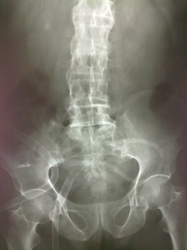
This X-Ray KUB, shows bilateral sacroilitis with diffuse syndesmophytic ankylosis (bamboo spine), ossification of spinal ligaments, joints and discs - suggestive of Ankylosing Spondilitis. And Radio opaque shadow in the Rt renal area due to Rt Renal Calculi.
In case of Ankylosing Spondylitis, a plain film must be noted for,
Sacroiliac joints:
Knees demonstrate uniform joint space narrowing with bony proliferation
Hands are generally involved asymmetrically, with smaller, shallower erosions and marginal periostitis.
ChestRadiographs of the lungs may demonstrate progressive fibrosis and bullous changes at the apices. These lesions may resemble TB infection and bullae may become infected.
Sacroiliac joints:
- a sacroiliitis is usually the first manifestation and is symmetrical and bilateral
- joints widen before they narrow
- subchondral erosions, sclerosis and proliferation on the iliac side of SI joints
- at endstage, the sacroiliac joint may be a thin line or not visible
- early spondylitis is characterized by small erosions at the corners of vertebral bodies with reactive sclerosis
- squaring of the vertebral body
- diffuse syndesmophyitic ankylosis can give a "bamboo spine" appearance
- interspinous ligament calcification can give a "dagger spine" appearance
- ossification of spinal ligaments, joints and discs.
- pseudoarthroses may form at fracture sites.
- enthesophyte formation from enthesopathy.
- Romanus lesions of the spine - shiny corner sign.
Knees demonstrate uniform joint space narrowing with bony proliferation
Hands are generally involved asymmetrically, with smaller, shallower erosions and marginal periostitis.
ChestRadiographs of the lungs may demonstrate progressive fibrosis and bullous changes at the apices. These lesions may resemble TB infection and bullae may become infected.
 RSS Feed
